inGenuyX provides biomedical engineering consulting services. Biomedical problems we analyze include, but are not limited to, the complex engineering environment in MRI scanners, where we can analyze not only the electromagnetic fields produced by all sets of coils but can also investigate on more secondary effects such as temperature changes in tissue and in the system.
We use advanced computational analysis and multiphysics simulation to answer your open biomedical engineering questions together with detailed human body models that accurately describe the physics in different tissues in the body.
Problems in the biomedical domain are almost always multiple physical phenomena that interact, such as vibrations, acoustics, and thermal interactions, as well as electromagnetic field interactions. Our multiphysics analysis can provide you with the tools that are needed to further your cost efficiency in your development process. We also offer broad practical experience in different biomedical areas to confirm the validity of these mathematical solutions.

Types of Physics
Electromagnetics
We can solve for electromagnetic fields within the body, such as incurred during an MRI scan or cellphone call. We can analyze specific metrics such as the Specific Absorption Rate (SAR), as well as electric and magnetic field exposure in different tissues.
Heat Transfer
The exposure to electric fields ultimately leads to tissue heating, which can be analyzed with the help of our coupled multiphysics methods. We can also take cooling incurred by blood flow into account using mathematical models built into the simulator.

Acoustics
Acoustic wave propagation in the human body is an important part of biomedical engineering, for both ultrasound and thermoacoustic technologies. In the latter case, we incur a problem that is coupled with both heat transfer and electromagnetic phenomena.
Structure and Material
Biomechanics and bone stresses can be analyzed as part of our biomedical modeling solutions.
Multiphysics
Each of the above represents a separate phenomena. These are, however, inevitably interconnected in nature, and the biomedical domain is likely one of the most important areas where different physics need to be analyzed in a coupled manner. Our service focuses on the interplay between these phenomena and can be customized to your needs.
Our Experience
Simone has longstanding experience in the field of biomedical engineering modeling as part of her postdoctoral and staff scientist research position at Stanford University. She has been working in the field of MRI engineering since 2012 and has previously worked at McGill University on an early breast cancer detection system using microwave imaging. Prior to her postdoctoral work, she focused on RF and wireless engineering in the field of passive mm-wave imaging and mixer design for mm-wave designs. Her undergraduate and graduate work was in mechatronics, which combines the fields of mechanical engineering, electrical engineering, and computer science. This multidisciplinary training makes Simone the ideal candidate to perform the modeling tasks for your multiphysics problems.
Selected biomedical projects
See an overview of Simone’s past biomedical work.
Ultra High-Field Magnetic Resonance Imaging
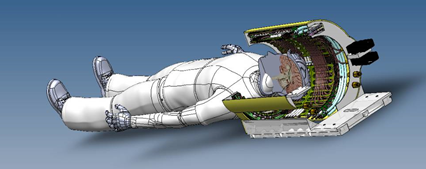
Magnetic Resonance Imaging (MRI) has emerged as one of the most powerful and informative diagnostic tools in modern medicine. While most clinical MR studies use magnetic field strengths of 1.5T or 3T, leading research is pushing these magnetic field strengths to 7T and beyond. These new ultra high-field (UHF) technologies promise images with higher spatial resolution, higher sensitivity to subtle change, and novel contrasts, which will improve our basic understanding of anatomy and physiology in both healthy tissue and disease. However, there are substantial hurdles to surmount before we will reap the promised benefits of UHF MRI in clinical applications. My research focuses on some of the major challenges faced in UHF MRI in engineering and multiphysics that are being researched to overcome these issues.
Selected publications
- S. A. Winkler, “MR engineering – A Handbook from the Bore’s Eye View,” Artech House Book Publishers – in application process.
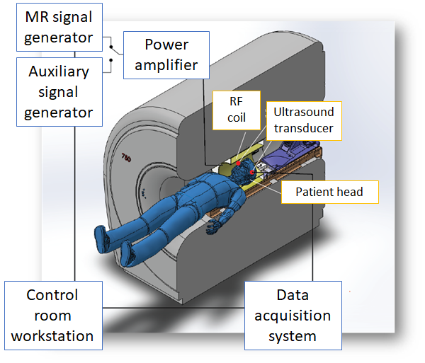
- S. A. Winkler, A. Alejski, T. Wade, C. McKenzie, B. K. Rutt, “On the Accurate Analysis of Vibroacoustics in Head Insert Gradient Coils,” Magnetic Resonance in Medicine, vol. 74, Iss. 4, Oct. 2017, pp. 1635-1645.
- S. A. Winkler, B. K. Rutt, P. Picot, M. Thornton, “Direct SAR Mapping by Thermoacoustic Imaging: A Feasibility Study,” Magnetic Resonance in Medicine, vol. 74, Iss. 4, Oct. 2017, pp. 1599-1606.
- Invited, Focused Issue: A. Winkler, F. Schmitt, H. Landes, J. De Bever, T. P. Wade, A. Alejski, B. K. Rutt, “Gradient and Shim Technologies for Ultra High Field MRI,” Neuroimage, Nov. 2016, pii: S1053-8119(16)30649-8.
- S. A. Winkler, B. K. Rutt, P. Picot, M. Thornton, “Method And System For Estimating The Specific Absorption Rate Of A Tissue Region Prior To A Magnetic Resonance Imaging Scan,” U.S. patent application 14/704,369, filed May 2015.
- S. A. Winkler, B. K. Rutt, “Practical Methods for Improved B1+-Homogeneity in 3T Breast Imaging,” Journal of Magnetic Resonance Imaging, Vol. 41, Iss. 4, pp. 992-9, Apr 2015. Epub 2014 Apr 10.
- Magna Cum Laude Award, Oral: S. A. Winkler, J. Stockmann, P. A. Warr, B. Keil, R. Watkins, L. L. Wald, B. K. Rutt, “Comparison of new element designs for combined RF-Shim arrays at 7T,” Proc. Int. Soc. Magn. Res. 2015, Toronto, ON, Canada, June 2015.
- 1 st place MR engineering award, Invited talk: S. A. Winkler, P. Picot, M. Thornton, B. K. Rutt, “Direct SAR Mapping by Thermoacoustic Imaging: Experimental Proof-of- Concept,” Proc. Int. Soc. Magn. Res. 2015, Toronto, ON, Canada, June 2015.
- S. A. Winkler et al., “Optimized Dielectric Shimming in High-Field Magnetic Resonance Imaging: A Theoretical Approach,” in Scattering by Aggregates on Surfaces, T. Wriedt, Y. Eremin, Eds. Berlin, Germany: epubli, 2014, pp. 70-73.
Early Breast Cancer Detection using Ultra-Wideband Microwave Imaging
A low-cost time-domain early breast cancer detection system based on microwave imaging, operating in the ultra-wideband (UWB) frequency range, has been developed and successfully employed for early detection of breast tumors. Highly realistic breast phantoms with irregular shape and different tissue contents have been constructed specifically for the purpose of tumor detection. A new transmitter system using specific UWB pulse shaping has been introduced leading to improved tumor detection performance. Latest results are shown and presented in comparison to prior experiments.
Selected publications
- Invited, Oral: A. Winkler, E. Porter, A. Santorelli, M. Coates, M. Popović, “Recent Progress in Ultra-Wideband Breast Cancer Detection,” Proc. Int. Symp. Ultra-Wideband, Syracuse, NY, USA, Sep. 2012, pp. 182-186.
- Oral: Porter, A. Santorelli, S. Winkler, M. Popović, “Time-Domain Microwave Cancer Screening: Optimized Pulse Shaping Applied to Realistically Shaped Breast Phantoms,” Proc. Int. Microwave Symp. 2012, Montreal, QC, Canada, Jun. 2012, pp. 1766-1769.
Images/Design by eyeCatchLight Photography and Shannon Merrell.
